Good documentation is the key to optimal coding and reimbursement for radiology procedures. By  including all of the essential elements in the radiology report, physicians give their coders all of the information they need to get the billing done most efficiently. But when the report lacks some required piece of data, the coders must contact the radiologist for clarification. At best, this slows down the billing process but, at worst, it leads to under-coding and therefore lower payment than is possible for the procedure.
A recent review of live radiology practice data by Healthcare Administrative Partners revealed that Venous Duplex Doppler exams regularly appear on the list of exams with insufficient reporting documentation. Following is a recommended checklist of elements that coders should use to review this exam:
Is the study complete?
A complete study includes bilateral upper or lower extremity imaging.
-
Complete bilateral lower extremity studies require documentation of all the following vessels: entire femoral vein, saphenous vein, and popliteal vein on both legs. The calf veins may also be included, but these are not required for a complete study.
-
Complete bilateral upper extremity studies require documentation of all the following vessels: subclavian, jugular, axillary, brachial, basilic and cephalic veins on both arms. Forearm veins may also be included, but these are not required for a complete study.
Is the study limited?
A limited study can be characterized by any of three different scenarios:
-
A bilateral study where not all the vessels required for a complete study are imaged;
-
A unilateral extremity study; or
-
A venous mapping for preoperative imaging (e.g. hemodialysis graft vessel mapping or vascular mapping for bypass grafts).
Are all three duplex techniques documented?
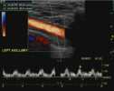
Gray scale imaging, spectral analysis, and color Doppler flow must all be used and documented on both complete and limited studies. If any one of these techniques is missing, a duplex imaging study was not performed and cannot be billed. It is necessary to document all techniques and provide interpretation for each of those techniques.
The Coding Committee of the Radiology Business Management Association (RBMA) provides coders with a helpful chart of “buzz words” that can be used when coding these exams:
Required Elements for Duplex Imaging |
||
|
DUPLEX = SPECTRAL ANALYSIS AND COLOR FLOW |
||
|
Element 1: |
Element 2: |
Element 3: |
|
B-Mode |
Acceleration |
Color Flow |
|
Diagnostic |
Bandwidth |
|
|
Gray Scale |
Mono, Bi or Tri |
|
|
Real-time Imaging |
Peak Systolic |
|
|
Two-Dimensional |
Phasicity |
|
|
|
Resistive |
|
|
|
Spectral |
|
|
|
Spectral Doppler |
|
|
|
Velocity |
|
|
|
Pulsed Doppler |
|
|
|
Pulsatility |
|
|
|
Waveform |
|
Radiologists can benefit by keeping these “buzz words” in mind when dictating their interpretation of Duplex Doppler exams. Documenting all of the required elements of these exams will allow your coders to bill for as complete a procedure as possible and you will be rewarded by achieving maximum reimbursement.
Healthcare Administrative Partners regularly reviews our clients’ reporting to provide recommendations to the radiologists for improvement of their documentation. Doing so is often the difference-maker in practices that realize maximum reimbursements. To determine if your practice performance is optimized, request our free coding assessment now.
Related articles:
The Impact of Coding Changes on Radiology Practices in 2015
Edited photo credit:
By JasonRobertYoungMD (Own work) [CC BY-SA 4.0 (http://creativecommons.org/licenses/by-sa/4.0)], via Wikimedia Commons


